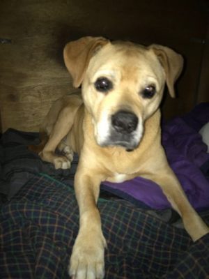
Bella, 11 year old Mastiff crossbreed
November 2019: Bella came back from a walk with a very swollen right side of her face and muzzle. My initial suspicion was that her fractured canine had become infected and caused an abscess. This was confirmed when I palpated the gum in the canine/premolar area. The swelling felt firm (like an abscess does) and was clearly causing pain. It was very clear that this must be an abscess. What to do …?
No luck at the vets.
We went to a vet in Truro and the vet in charge told us that this must be an allergic reaction… “probably a bee sting…” gave a shot of Antihistamines and told me to get some over the counter anti-allergic tablets. When I ask about the close proximity of a broken canine which might be infected, I was told: ” unlikely that they are the cause but they should be removed anyway…”
The next day, Bella still looking pretty bad, I went to get a 2nd opinion at another vet. After a very brief look at the dog, the vet concluded that this must be an allergic reaction and gave her a Steroid injection….. When I mentioned the broken canine I was offered a “dental appointment” 2 weeks later to remove both canines.
A canine is between 50 and 60mm long and the removal of this tooth is a surgical procedure. There is another option.
I have treated similar cases in 2007 and 2008 successfully.
Diagnosis and Treatment
With the help and supervision of Chris Gardner at St Clements Vets in Truro I could carry out a root canal treatment on both of Bella’s fractured canines.

After the second day the abscess burst with pus coming out (fistula). At that stage, the diagnosis of a subperiosteal abscess originating from the fractured right canine seems to be more likely than an allergic reaction.
Palpating a subperiosteal swelling, that means a swelling underneath or within the bone, gives a distinct feeling of firmness and tension under the surface, This is very different from a subcutenous swelling caused by an allergic reaction.
Unfortunately many vets still remove teeth which can be saved. Even though other treatment options are common practice in more advanced veterinary practices. The canine tooth of a dod can be 60mm long and to remove this tooth requires some serious osteotomy (removal of bone to uncover the root).
Treatment
Step 1
Pre-OP X-ray shows a discrete sinus around the canine root.
That is an indication of an infection.

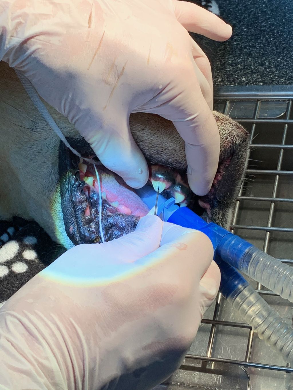
Inserting a root canal instrument in the open and infected canal. Confirmation of an infected canal.

Step 2
X-ray with an instrument in the canal to confirm the length of the root canal. Perfect preparation of canal.

Finishing the Root Canal treatment using the Lateral Condensation Technique.
Step 3
The control X-ray shows the finished Root Canal Fillings on the right and left canine.

Impression taking for 2 crowns using a human impression tray and silicon material.

Step 4
Crown/Root cap fitted on the right canine. The technician made it too short but it’s still effective to protect the tooth.

Because the canines were very short, we fitted very short crowns to avoid stress fractures.

Waking up.
What a strange dream …

